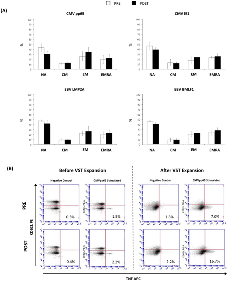
Figure 4. Panel A shows proportions (%) of naïve (NA), central memory (CM), effector memory (EM) and RA+ EM (EMRA) cells among CD8+ T-cells expressing TNF-α in response to viral peptide stimulation at Day 8.
No significant differences in the composition of CD8+ T-cell subsets were found between the cells expanded before (PRE) or after (POST) exercise (p > 0.05). Panel B shows representative flow cytometry plots from the TNF-α intracellular cytokine staining assays following CMVpp65 stimulation or control (no peptides) of PBMCs obtained before (PRE) or after (POST) exercise prior to and after ex vivo expansion. Values in the lower right quadrants are expressed as the percentage of all CD8+ T-cells. The phenotypic characteristics of the peptide-responsive VSTs cells (Panel A) were determined through the co-expression of CD62L and CD45RA on the CD8+/TNF-α+ events by flow cytometry.

