
Figure 2.
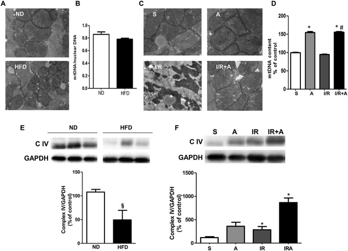
Prevention of mitochondrial damage by apelin‐13 in acute phase of myocardial I/R in obese mice. (A) Typical electron micrographs (original magnifications ×10000) from ND‐ and HFD‐fed mice. (B) Quantitative RT‐PCR analysis of mtDNA content in hearts from ND‐ and HFD‐fed mice. (c) Scanning electron microscope images (original magnification ×10000) in mice treated with vehicle or apelin‐13, A, after 24 h of sham‐operation, S, or I/R. (D) Effect of apelin‐13 treatment on mtDNA content in hearts from HFD‐fed mice subjected to I/R. (E) Western blot analysis of the myocardial OXPHOS complex IV in ND‐ and HFD‐fed mice. (F) Effect of apelin‐13 treatment on expression of OXPHOS complex IV in hearts from HFD‐fed mice subjected to I/R. *, P < 0.05 versus S; #, P < 0.05 versus I/R; §, P < 0.05 versus ND.