Figure 4.

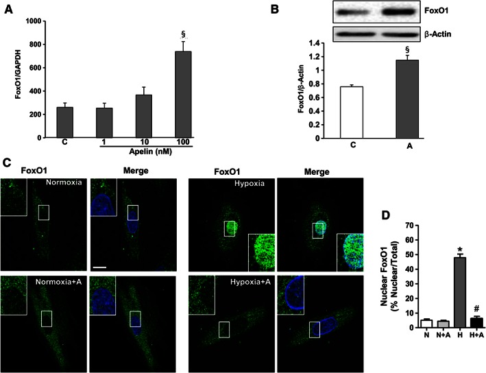
FoxO1 activity is regulated by apelin in H9C2 cardiomyoblasts. (A) Dose‐dependent effect of apelin‐13 on FoxO1 expression levels was assessed by quantitative RT‐PCR in H9C2 cardiomyoblasts treated or not with apelin (1–100 nM) for 24 h. (B) H9C2 cells were treated with apelin (A, 100 nM), and cell lysates were probed with anti‐FoxO1 and anti‐β actin antibodies. FoxO1 protein expression levels were quantified by densitometry and normalized against β actin. (C) Representative confocal images of H9C2 cells pretreated or not with apelin for 15 min (A, 100 nM), submitted to hypoxia, H, or normoxia, N, for 2 h and stained for FoxO1 antibody. FoxO1 proteins are shown in green; nuclei were stained with DAPI (in blue). Bar is 10 μm. (D) Quantification of FoxO1 nuclear translocation is expressed as the fluorescence intensity in the nucleus normalized to the total fluorescence intensity in the cell. *, P < 0.05 versus N; #, P < 0.05 versus H; §, P < 0.05 versus C.
