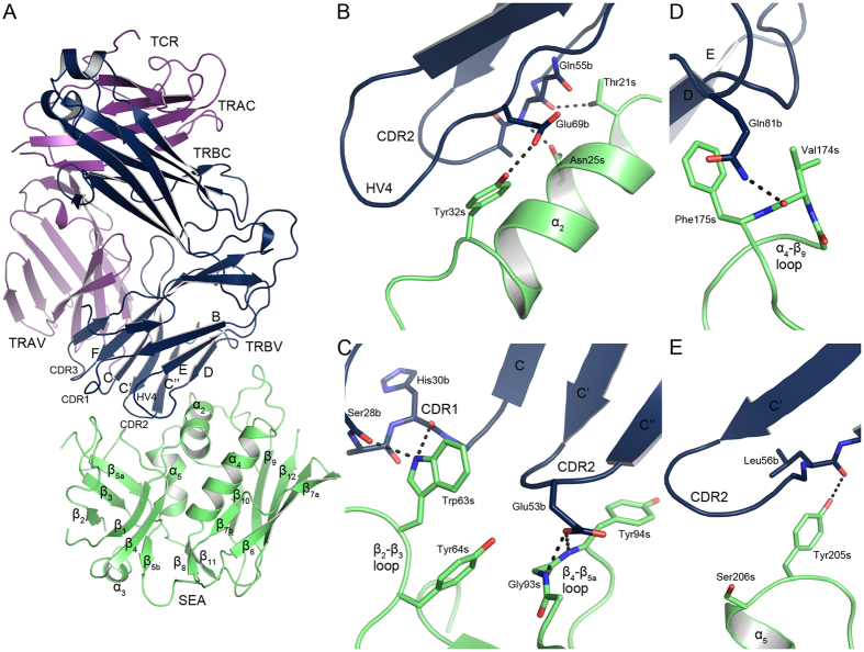
Figure 1. Structure of the SEAF47A-TCR complex.
(A) Overall structure of SEAF47A-TCR, with SEA shown in green, and TCR in purple and blue for the α- and β-chain, respectively. (B) Close-up view of the SEA α2-helix and contacting residues in TCR, (C) the hydrophobic patch, consisting of the β2-β3 and β4-β5a loops, (D) the α4-β9 loop, (E) and the upper part of the α5-helix. Hydrogen bonds are marked as black dotted lines and residues designated as “s” for SAg and “b” for TRBV7-9.

