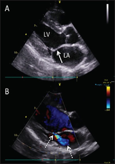
Figure 1.

Parasternal long-axis image of a typical case of latent, definite RHD. (a) 2D image highlights the thickened mitral valve leaflet with a characteristic “dog-leg” deformity (solid arrow) (b) Color Doppler image shows characteristic posteriorly-directed mild mitral regurgitation (dashed arrows)
