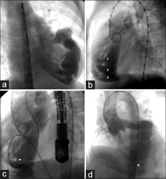
Figure 1.

Catheter intervention. (a and b) Aortogram 39° showing a grossly dilated left anterior descending coronary artery. Large communication is visible between the coronary artery and the right ventricle via three orifices (labeled 1, 2, and 3 in panel B). (c) Aortogram, lateral projection. A balloon (the white dot) was used to estimate the size of the orifice. The transesophageal echocardiogram probe is visible at the right. (d) Aortogram, left anterior oblique projection. The white dot indicates the ADO device that was placed in the lower orifice
