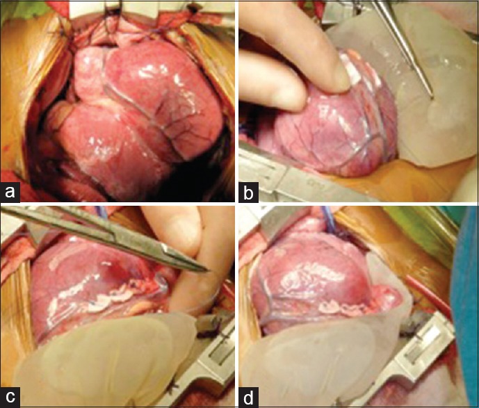
Figure 2.

The surgical intervention. (a) The left pericardium was retracted using several sutures. (b) The area surrounding the left anterior descending artery was stabilized using a surgical glove filled with sterile serum placed under the heart. (c) Using data obtained from the transesophageal echocardiograph, the orifices were located. The cardiologist guided the surgeon, who then began to suture below the left anterior descending artery. The sutures were backed with Teflon patches. (d) The procedure was completed after six sutures were placed under the artery
