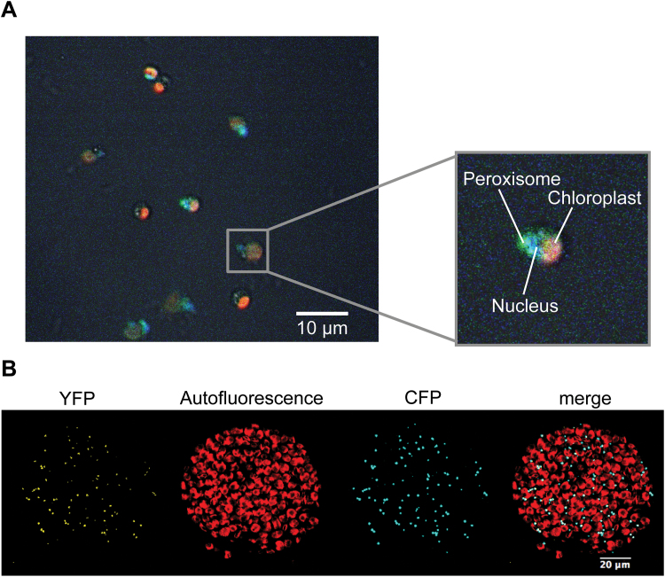
Fig. 3.
Subcellular localization studies of CmGOX. (A) Localization of CmGOX in C. merolae. Red: chloroplast autofluorescence; blue: DAPI-stained nucleus; green: YFP signal from YFP::CmGOX construct. C. merolae cells were transformed 24h before microscopic analysis. (B) Localization of CmGOX in tobacco protoplasts. From left to right: YFP signal of YFP::CmGOX construct, chlorophyll autofluorescence, CFP signal as peroxisomal marker (CFP::PTS1), and merge of all three pictures. Microscopic analyses were performed with protoplasts isolated from transiently transformed N. benthamiana leaves.

