Fig. 1.

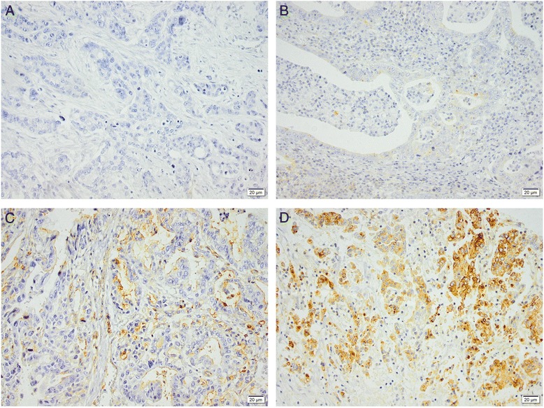
Sample immunohistochemical images of IFITM1 staining in gastroesophageal adenocarcinoma primary tumors with (a) negative, (b) weak, (c) moderate, and (d) strong staining of tumor cells. Magnification x 20

Sample immunohistochemical images of IFITM1 staining in gastroesophageal adenocarcinoma primary tumors with (a) negative, (b) weak, (c) moderate, and (d) strong staining of tumor cells. Magnification x 20