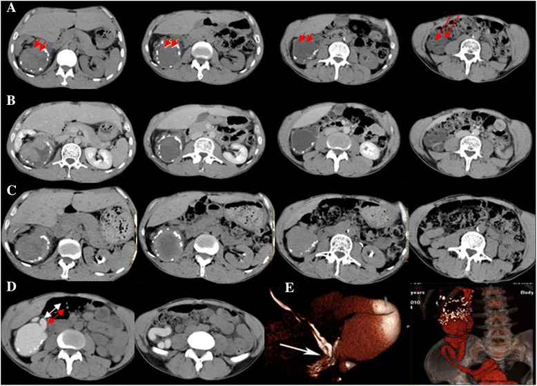
Fig. 1.

Case 1. A 56-year-old male patient with right side duplex kidney. Unenhanced supine axial CT (a) shows a solitary round, iso-dense, soft-tissue mass with a clear boundary (short arrow). The irregularly annular calcified shadow around the mass was equivalent to multiple annular low-density shadows in the ileocecal junction (long arrow). The mass was not clearly intensified after contrast enhancement (b) and one-hour delayed (c) CTU scanning. Interestingly, it was obviously strengthened after 18-h delay (d), confirming duplex kidney. Furthermore, we found a band of high-density shadows that was confirmed to be duplicated ureters (double-headed arrow). Reformatted 3D CTU after segmentation of bone structures also showed the entire course of the dilated ureters (e)
