
Fig. 4.
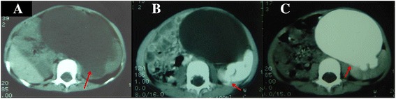
Case 4. CT scans obtained in a 2.5-year-old girl with left duplex ureters and congenital megalo-ureter. Non-enhanced scan obtained at the level of the inferior-middle pole of the left kidney shows a huge round-like area of low density (red arrow) (a). Contrast-enhanced scans were obtained at the same level as in (a), this area showed no enhancement on (b) the 26-h scan, and the tortuous contrast agent can be seen retroperitoneally (red arrow) (b). The huge round-like area of low density was intensified on the 48-h scan (red arrow) (c)