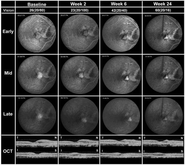
FIGURE 2.
Patient 5, a 37-year-old Caucasian woman with CNV attributable to angioid streaks. Studies obtained at baseline and 2, 4, and 24 weeks after the first infusion are shown including BCVA in ETDRS letters read at 4 meters and Snellen equivalent, frames from early, mid, and late phases of FA, and horizontal and vertical cross-sections of OCT scans labeled T-N (temporal-nasal) and I-S (inferior-superior), respectively. At the primary endpoint of 24 weeks after the first infusion, the patient showed improved BCVA, reduced CNV lesion size, resolution of leakage, and resolution of subretinal and intraretinal fluid in the macula.

