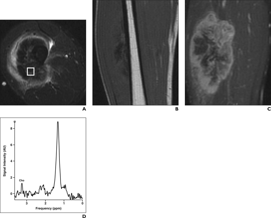
Figure 3.
31-year-old man with high-grade osteosarcoma of right thigh.
A, Axial STIR fast spin-echo MR image (TR/TE, 4650/47) shows large heterogeneous mass in anterolateral right thigh with surrounding soft-tissue edema.
B, Coronal T1-weighted spin-echo MR image (TR/TE, 550/11) shows low-signal-intensity mass.
C, Coronal fat-saturated dynamic contrast-enhanced fast spin-echo MR image (TR/TE, 600/10) acquired 40 seconds after contrast injection shows heterogeneous enhancement with central areas of necrosis.
D, Single-voxel MR spectroscopic map shows discrete choline (Cho) peak at 3.2 ppm. Signal-to-noise ratio is 5.4.

