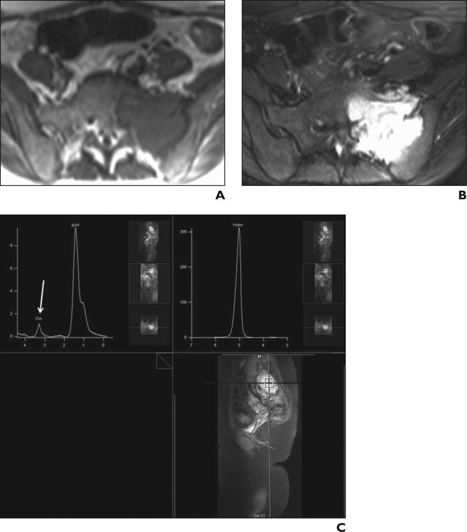
Figure 5.
25-year-old woman with Ewing sarcoma of left sacrum.
A, Axial T1-weighted spin-echo MR image (TR/TE, 690/15) shows left sacral mass as low-signal-intensity lesion at sacroiliac joint.
B, Axial fat-suppressed fast spin-echo T2-weighted MR image (TR/TE, 2886/100) shows involvement of neural foramina, crossing of sacroiliac joint, and associated soft-tissue component in anterior aspect. Although conventional MRI findings suggested malignant features, diagnosis of giant cell tumor was entertained in light of some characteristic features.
C, Single-voxel proton MR spectroscopic map obtained within sacral mass with weak water suppression (top left) shows discrete choline peak (arrow). Relative signal-to-noise ratio is 3.6. Absolute choline concentration was calculated to be 2.9 mmol/kg, suggesting malignancy in this pathologically proven Ewing sarcoma. Upper right image is not water suppressed.

