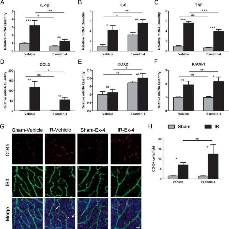
Figure 2.
Exendin-4 inhibits the induction of the expression of IR-responsive genes associated with inflammation without affecting the number of CD45-positive cells. Rats were treated with Ex-4 (10 μg/kg) twice daily with two initial administrations prior to ischemia, and injections every 12 hours for the next 48 hours during the reperfusion period. Nontreated animals received saline vehicle injections. One eye of each animal was subjected to retinal ischemia for 45 minutes, followed by natural reperfusion. The contralateral eyes were subjected to needle puncture and served as sham controls. Total RNA was isolated from the retinas and the relative levels of mRNA of (A) IL-1β, (B) IL-6, (C) TNF, (D) CCL2, (E) COX2, and (F) ICAM-1 were determined by duplex qRT-PCR with β-actin serving as control. Data are expressed as mean ± SEM (n = 8 retinas per group). (G) Retinas were isolated and stained with antibodies to CD45 (red), isolectin B4 (IB4, green), and Hoechst nuclear stain (blue) and then flat mounted. The majority of CD45-positive cells are present in the perivascular region (arrows) with few cells positively staining inside the vessels (arrowhead). Scale bar: 25 μm. (H) We quantified CD45-positive cells in each retina from each group. Data are presented as CD45-positive cells per field and represent the mean ± SEM (n = 8 retinas per group). *P < 0.05, **P < 0.01, ***P < 0.001. One-way ANOVA followed by Bonferroni's post hoc test.

