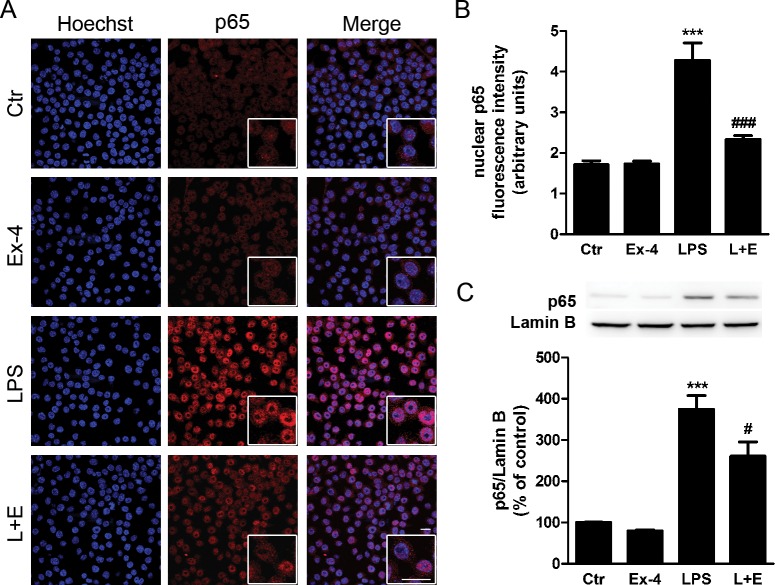
Figure 6.
Exendin-4 inhibits the nuclear accumulation of the NF-κB p65 subunit in LPS-stimulated BV2 microglial cells. (A) We treated BV2 cells with Ex-4 (100 nmol/L) 1 hour prior to the LPS stimulus (100 ng/mL for 30 minutes). Subcellular localization of p65 subunit (red) was evaluated by immunocytochemistry. Hoechst staining (blue) was used to visualize nuclei. Scale bar: 20 μm. (B) Quantification of nuclear fluorescence intensity for p65 immunoreactivity in BV2 cells. Data are presented as arbitrary fluorescence units and represent the mean ± SEM (n = 3). (C) We stimulated BV2 cells as described in (A). Subcellular fractionation was performed and nuclear extracts were separated by SDS-PAGE and immunoblotted with anti-p65 antibody. Lamin B was used as a loading control. Quantification of p65 protein levels was performed by densitometric analysis. Data are presented as mean ± SEM (n = 5). ***P < 0.001 versus control. #P < 0.05, ###P < 0.001 versus LPS. One-way ANOVA followed by Bonferroni's post hoc test.

