Fig. 1.
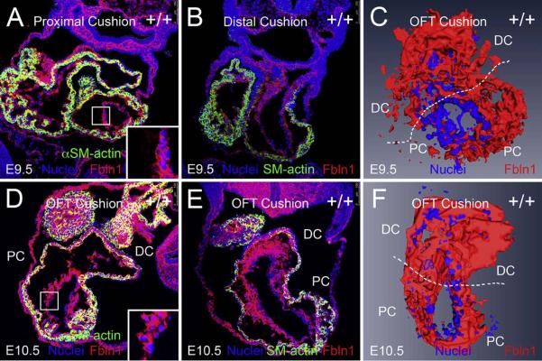
Fibulin-1 is present in the proximal and distal OFT cushions at E9.5 and E10.5. (A and B) show cryosections (10 μm) of wild type E9.5 OFT cushions. (A) shows E9.5 proximal cushions and (B) shows E9.5 distal cushions immunolabeled using anti-Fbln1 (red) and anti-αSMA (green). Nuclei were stained using Hoechst (blue). (D and E) show cryosections (10 μm) of wild type E10.5 OFT cushions. (D) shows E10.5 proximal and (E) shows E10.5 distal cushions immunolabeled using anti-Fbln1 (red) and anti-αSMA (green). Nuclei were stained using Hoechst (blue). Inset boxes in A and D show portions of the proximal OFT at higher magnification. Lumen region in insets is to the right; Fbln1 (red) is seen on the inner surface and in close association with the endocardium (blue nuclei). Shown in A–B and D–E are representative images of OFT cushions from 2 different wild type E9.5-E10.5 embryos. PC, proximal cushions; DC, distal cushions. All sections are transverse. (C and F) AMIRA-based 3D reconstructions of OFT, endothelial and EMT-derived mesenchymal cells (both Hoechst labeled, blue) and adjacent Fbln1 (red) in wild type E9.5-E10.5 proximal and distal OFT. 3D reconstruction regions are limited to the EMT-derived sections of E9.5-E10.5 proximal and distal OFT cushions. PC, proximal OFT cushions; DC, distal OFT cushions.

