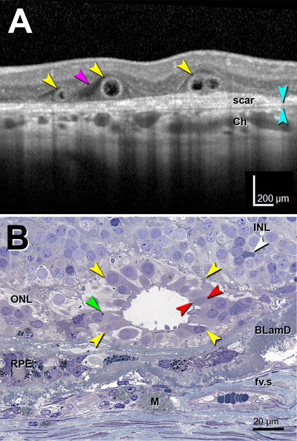
Figure 1.
Optical coherence tomography imaging and histology of outer retinal tubulation. Outer retinal tubulation cross-sections (yellow arrowheads). (A) Representative SD-OCT B-scan of three ORT cross-sections from TCS1 volume from an 81-year-old woman with neovascular AMD. Two Closed ORTs on the left, and a Branching ORT on the right. Hyporeflective wedge38 (pink arrowhead); Bruch's membrane (cyan arrowheads). (B) High-resolution histology section of degenerate cones in ORT, at 1.5 mm from the fovea from a different 81-year-old woman with neovascular AMD. Cone lipofuscin (green arrowhead); mitochondria in outer fiber39 (red arrowheads); Müller cell body (white arrowhead). BLamD, basal laminar deposit; Ch, choroid; fv.s, fibrovascular scar; INL, inner nuclear layer; M, lipid-containing macrophage; RPE, entombed and melanotic retinal pigment epithelium.40

