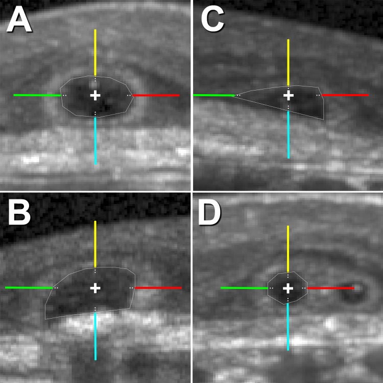
Figure 2.
Outer retinal tubulation types in cross-section for intensity analysis. In cross-sections, ORT is identified as Closed (A), Open (B), Forming (C), or Branching (D). For the intensity analysis, hyperreflective ORT bands in cross-sections were sampled at 0° (red), 90° (yellow), 180° (green), and 270° (cyan). Raw (linear) SD-OCT intensity (ranging from 0–1 in a 32-bit image) by ORT type and angle was sampled at 5-μm steps from the inner aspect of ORT band along the colored lines, and the average first maximum intensity value is reported in Figure 5 and Supplementary Figure S2 for each SD-OCT volume. Each line of the cross in the centroid of ORT cross-sections is 20 μm long.

