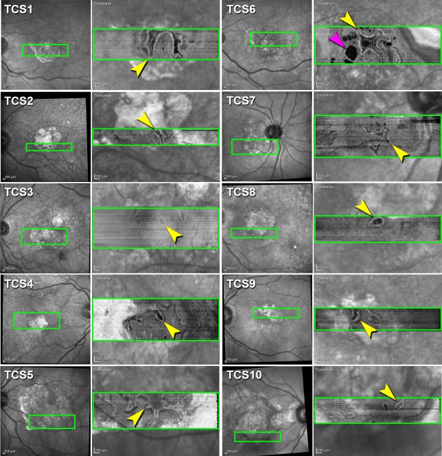
Figure 3.
Outer retinal tubulation varies in size and shape by SD-OCT en face reconstruction. Infrared reflectance (IR) images of 10 eyes (first and third columns). Green box is location of en face transverse reconstruction image from each of 10 SD-OCT volumes (Heidelberg Spectralis) of nine patients with neovascular AMD showing ORT network (yellow arrowheads) overlaid on IR image (second and fourth columns). Cyst (pink arrowhead).

