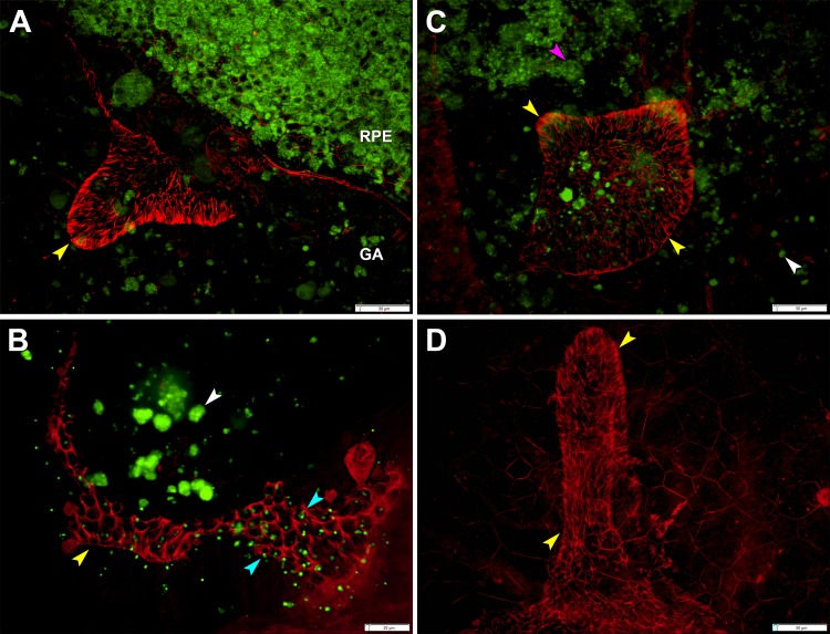
Figure 6.
Outer retinal tubulation in a flatmount retina. (A) Outer retinal tubulation (yellow arrowheads) marked by ELM cytoskeleton meshwork at the border of geographic atrophy (GA) 5.5 mm from the fovea. (B) Autofluorescent cone lipofuscin granules (cyan arrowheads) in ELM meshwork through which ORT cones point into lumen at 2.6 mm from fovea. Retinal pigment epithelium granule aggregates (white arrowheads). (C) Outer retinal tubulation overlying dissociated RPE25 at 5.1 mm from the fovea. Spherical RPE cell in the process of degranulating (pink arrowhead). (D) Outer retinal tubulation of different morphology at 4 mm from fovea; RPE autofuorescence is not shown. Tissue from 86-year-old woman with neovascular AMD, labeled with Alexa 647 Phalloidin. Lipofuscin is shown by 488-nm autofluorescence. (A–D) were used, with others, to calculate cross-sectional cone area (49.1 ± 7.9 μm2), center-to-center cone spacing (7.5 ± 0.6 μm), and cone density (20,351 cones/mm2) in ORT.

