
Figure 1.
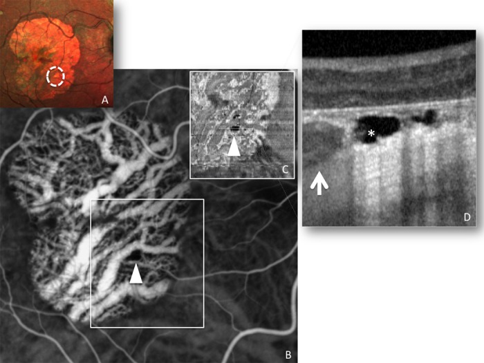
MultiColor imaging, ICGA en face optical coherence tomography (OCT), and B-scan OCT of choroidal caverns. MultiColor imaging (A) shows refractile intense hyperreflective material in correspondence of the choroidal caverns (dotted circles). Indocyanine green angiography (B) and en face OCT (C) of choroidal caverns (asterisk) reveals that the areas occupied by these cavities are devoid of significant blood flow (arrowheads). Optical coherence tomography B-scan (D) shows the choroidal caverns as gaping hyporeflective cavities in the choroid (both Sattler and Haller layers, with relative preservation of the choriocapillaris), well distinguishable from choroidal vessels, which are slightly hyperreflective due to the presence of blood, with characteristic hyperreflective border due to the vessel wall (arrow). The cavities in choroidal caverns are typically empty, angular, with punctate hyperreflectivities internally (asterisk).