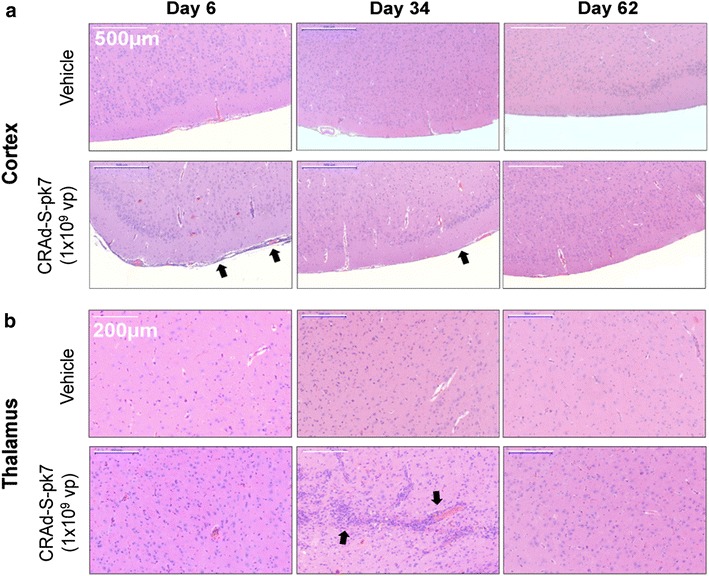
Fig. 7.

Histological analysis of samples associated with CRAd-S-pk7 adenoviral vector delivery over time. Assessment of histopathological changes on CNS tissues associated with adenoviral vector treatment. Vector high dose group (1 × 10⁹) is shown. In a minor amounts of meningitis perivascular inflammation were detected in the treatment groups, which subsided by 62 days post treatment. In b minor inflammation of the thalamus and its associated vasculature occurred in select animals; which also subsided by 62 days post treatment. Quantification of events is outlined in Additional file 1: Table S2. For histological examination, we used 10 hamsters per group per time point
