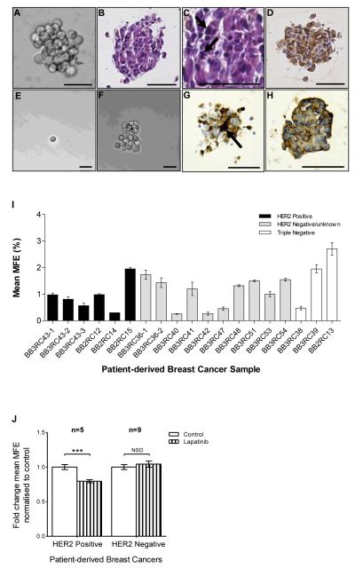
Figure 1. Mammosphere formation from patient-derived breast cancers and effects of EGFR/HER2 inhibition.
A Typical bright-field photomicrograph of a mammosphere (>60μm in diameter) grown from single cells plated at 500 cells per cm2 after 7 days in non-adherent culture. B-D Photomicrographs of 3μm sections of a FFPE patient-derived mammosphere showing B) H&E staining C) pleomorphic nuclei (arrows) and D) pancytokeratin expression (brown). Mammospheres generated after 7 days were dissociated into single cells and re-plated at 1 cell per well; E) Bright-field photomicrograph of a single cell re-plated in a 96 well plate; F) a mammosphere regenerated from a single cell plated per well after 10 days in non-adherent culture. G and H Photomicrographs of 3μm sections of a FFPE patient-derived mammosphere showing G) ALDH1 expression (brown staining), arrow shows intense expression in a single cell and H) CXCR1 expression (brown staining). A-H Scale bars = 50μm. I MFE of 19 independent patient-derived breast cancers. There was no significant difference in MFE between HER2 positive, HER2 negative and triple negative cancers. Columns, mean; bars, SEM of triplicate observations. J MFE of HER2 positive (n=5) and HER2 negative (n=9) cancers after 7 days in non-adherent culture with/without lapatinib (1μM). Controls were treated with vehicle (0.01% DMSO). Columns, fold change in MFE normalised to control; bars, SEM. ***, P <0.001; NSD, no significant difference.

