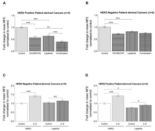
Figure 3. Interaction between CXCR1/2 and HER2 signalling in mammosphere formation.
HER2 positive (n=4) and HER2 negative (n=9) patient-derived breast cancer cells were plated out in non-adherent culture conditions. A and B show the effect of SCH563705 (100nM), lapatinib (1μM) or a combination of lapatinib and SCH563705 at the stated respective doses on MFE in A) HER2 positive B) and HER2 negative breast cancers. All conditions, including control were supplemented with recombinant IL-8 (100ng/ml). C and D show the effect of IL-8 (100ng/ml), lapatinib (1μM) or lapatinib and IL-8 at the stated respective doses on MFE in C) HER2 negative and D) HER2 positive breast cancers. Controls were treated with vehicle. A-D Columns, fold change in MFE normalised to control; bars, SEM. *, P < 0.05; **, P < 0.01; ***, P < 0.001; NSD, no significant difference.

