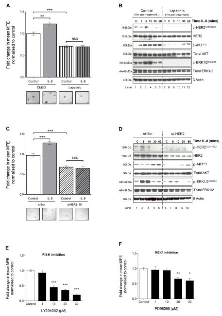
Figure 4. CXCR1/2-mediated effects on mammosphere formation and downstream signalling pathways are dependent on HER2 phosphorylation.
A MCF7/HER2-18 cells were plated out in non-adherent culture conditions and treated with either IL-8 (100ng/ml), lapatinib (10μM) or lapatinib and IL-8 at the stated respective doses. Controls were treated with vehicle. Graph shows the effect of treatments on MFE. Inserts show representative bright-field photomicrographs of mammospheres cultured under each condition; scale bars = 100μm. B MCF7/HER2-18 cells were plated out in adherent culture conditions and pre-treated with either vehicle control (lanes 1-6) or lapatinib (10μM, lanes 7-12) for 1 hour prior to stimulation with IL-8 (100ng/ml) for 2, 5, 10, 30 and 60 minutes. Controls (C) were treated with vehicle for 10 minutes. Immunoblots show the effect of IL-8 on phosphorylation of HER2, AKT and ERK1/2. C MCF7/HER2-18 cells transfected with either scramble control siRNA (siScr), or siRNA to HER2 (siHER2-15) were plated out in non-adherent culture conditions and treated with either IL-8 (100ng/ml) or vehicle control. Graph shows the effect of these treatments on MFE. Inserts show representative bright-field photomicrographs of mammospheres cultured under each condition; scale bars = 100μm. D MCF7/HER2-18 cells were transfected with either siScr (lanes 1-5), or siHER2-15 (lanes 6-10). After 24 hours, cells were serum starved for 48 hours and then stimulated with IL-8 (100ng/ml) for 5, 10, 30 and 60 minutes. Controls (C) were treated with vehicle for 10 minutes. Immunoblots show the effect of IL-8 on phosphorylation of HER2, AKT and ERK1/2. E and F MCF7/HER2-18 cells were plated out in non-adherent culture conditions and treated with increasing doses of LY294002, a PI3-K inhibitor (E) and PD98059, a MEK1 inhibitor (F). Graphs show the effect of treatments on MFE. A, C, E and F Columns, fold change in MFE normalised to control; bars, SEM, n=3 independent experiments. **, P < 0.01; ***, P < 0.001; NSD, no significant difference. B and D blots are representative of 3 independent experiments. β-actin was used as a loading control; molecular weight for each protein is shown.

