
Figure 8.
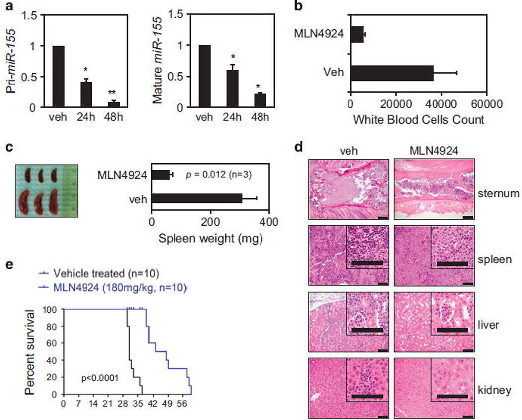
Antileukemic activity of MLN4924 in a human AML xenograft model. (a) NOD/SCID γ (NSG) mice were transplanted intravenously with MV4-11 cells. After the engraftment, splenic cells from the leukemic mice were injected into secondary recipient NSG mice. One week later, 10 mice were treated with 180 mg/kg of MLN4924 or vehicle control. Pri-miR (left) and mature miR-155 (right) expression levels in the peripheral blood of xenografted mice were measured by real-time PCR, as early as 24 and 48 h after the first dose of MLN4924 and compared with vehicle control (veh). Results of mRNA expression are shown as relative fold change after normalization to 18 S and U44, respectively, and 2 Ct calculations. Error bars represent s.d. (P <0.01 for 48 h) based on triplicate readings of average of two separate experiments. (b) Average white blood cell count (µl/ml) taken from three mice representing each group and killed 21 days after the first injection of MLN4924 or vehicle control (veh). (c) Spleen pictures of three representative cases (MLN4924 n = 3; vehicle n = 3) at 21 days. Spleens were weighed and results are shown as bar graph representing the average of three in each group. P-value obtained using t-test is shown. (d) Hematoxylin and eosin staining of sections from sternum, spleen and liver of xenografted mice treated with vehicle or MLN4924 at 21 days. (e) Survival plot of xenografted mice after the treatments with MLN4924 at 180 mg/kg (n = 10) or vehicle control (veh; n = 10). Survival comparison was made using the log-rank test.