
Fig 1. Identification TNC in semen and CVL.
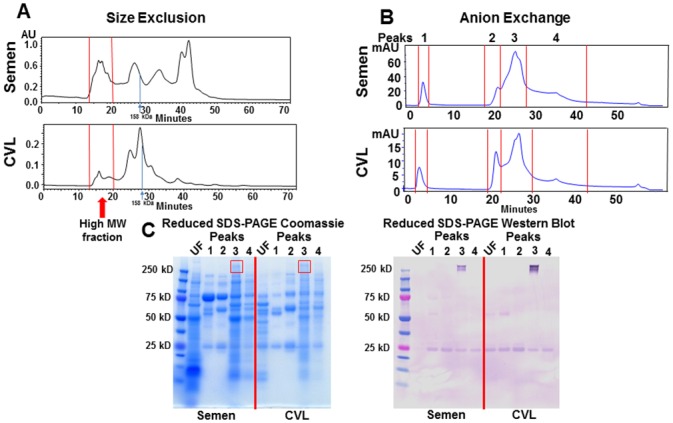
(A) Pooled semen and CVL was separated by size (>500 kDa) and charge (B) by high performance liquid chromatography (HPLC). (C) Unfractionated (UF, diluted 1:10) and charge-separated, high molecular weight (MW) semen and CVL protein peaks were then run on a reduced SDS-PAGE gel and Coomassie stained. TNC was detected with an anti-TNC monoclonal antibody. The 250 kD band staining on anti-TNC western blot was confirmed to be TNC by LC/MS.