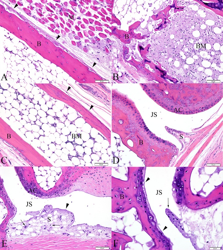
Fig 7. Metatarsal bones and tarsal joints from IRF 3/7 -/- -/- mice.
(A) 4 DPI, periosteal necrosis of the metatarsal bone (arrowheads). The periosteum is slightly expanded by fibrin and cellular debris. (B) 4 DPI, distal aspect of metatarsal bone exhibiting extensive bone marrow necrosis. Normal bone marrow is replaced by fibrin, cellular debris, and necrotic and apoptotic cells. (C) Metatarsal bone from a PBS-inoculated control mouse demonstrating normal bone marrow and periosteum (arrowheads). (D) Synovium and articular cartilage from a normal PBS-inoculated control mouse. (E) 5DPI, fibrinous synovitis exhibiting loss of synoviocytes, accumulation of fibrin along the intima and within the superficial subintima (arrowhead), and infiltration of the subintima by few leukocytes (arrows). (F) 6 DPI, cartilage necrosis (bounded by arrowheads) is apparent in the articular cartilage of the metatarsal bone, adjacent to a region of fibrinous synovitis (arrow). (B = bone, M = skeletal muscle, BM = bone marrow, S = synovium, JS = joint space, AC = articular cartilage)

