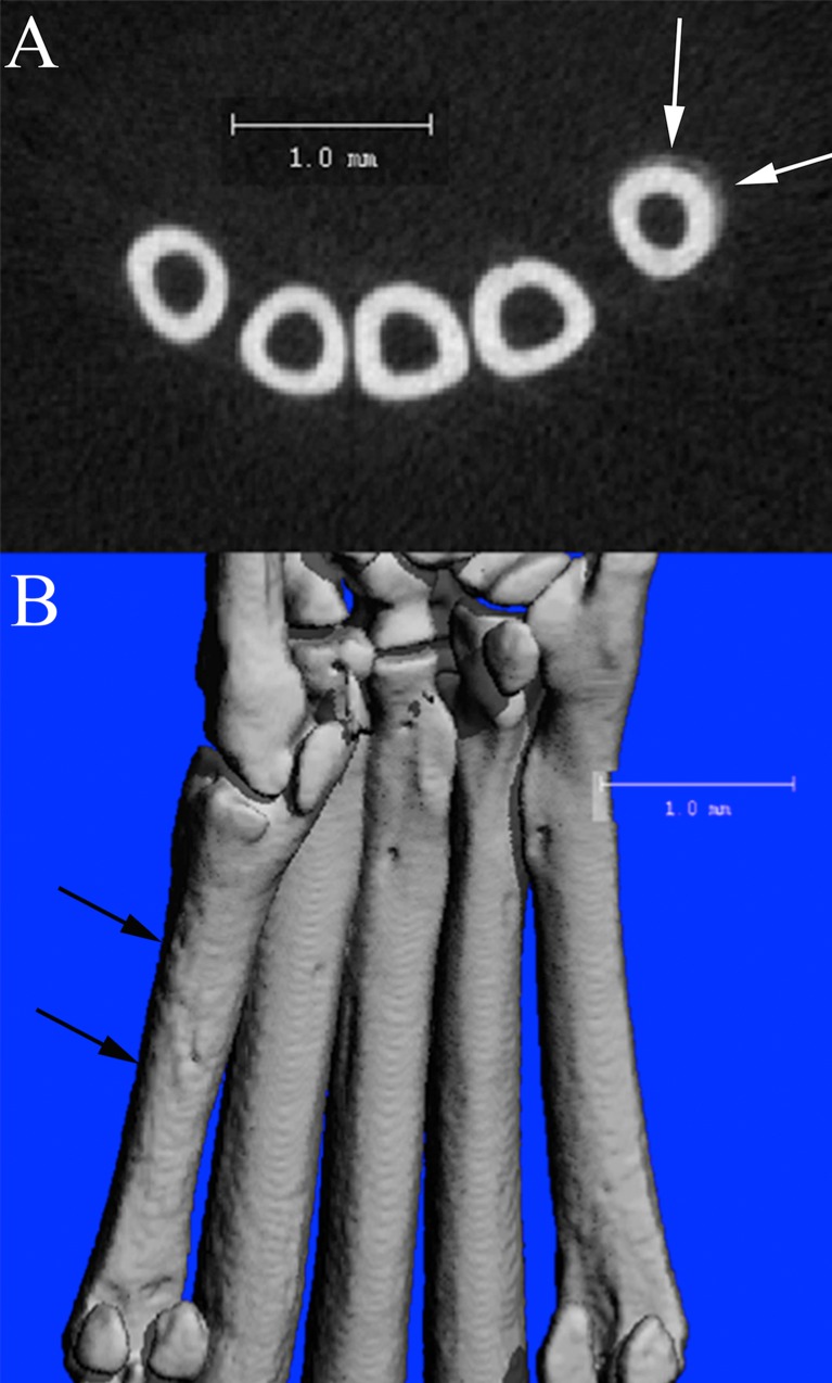
Fig 10. μCT scans of a CHIKV infected C57BL/6 mouse at 21 DPI.
(A) 2D slice from μCT scan of the mid-metatarsal region demonstrating region of periosteal bone proliferation (arrows) on first metatarsal bone (Mt1). (B) Corresponding 3D reconstruction of the plantar metatarsal region of the same mouse, demonstrating the roughened periosteal surface on Mt1 (arrows) in the region of the proliferative lesion identified in A.

