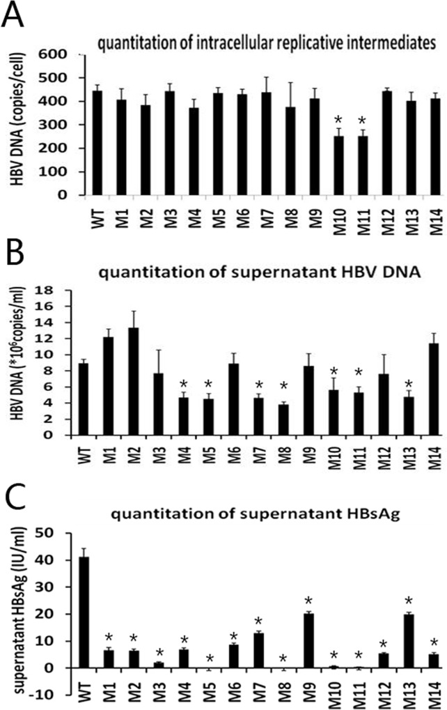
Fig 3.
Quantitation of intracellular replicative intermediates (A), supernatant HBV DNA (B), and supernatant HBsAg (C). WT, wild-type; M1, sQ129N; M2, s131−133TSM→NST; M3, sI126V; M4, sG145R; M5, sI126V+sG145R; M6, s115−116 “INGTST” insertion; M7, s115−116 “INGTST” insertion+sG145R; M8, s122−123 “KSTGLCK” insertion+sQ129N; M9, s126−127 “RPCMNCTI” insertion; M10, nt 3014−3198 deletion; M11, nt 3046−3177 deletion; M12, preS2 initiation codon M→I+s131+133TSM→NST; M13, nt 2848−2862 deletion+preS2 initiation codon M→I; M14, nt 3115−3123 deletion+sQ129N (* P < 0.05, mutant vs. WT).

