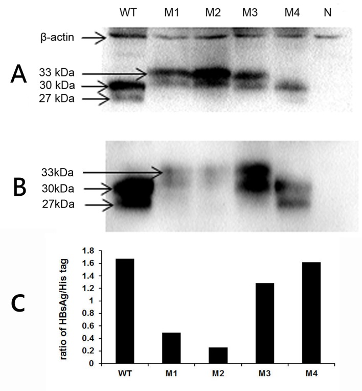
Fig 4. Western blot analysis of His-tagged HBsAg.
His-tagged target proteins were detected using anti-His tag monoclonal antibody (A) or anti-HB monoclonal antibody (B). A relative densitometry analysis of the bands was performed using a Tanon gel image system (C). WT, wild-type; M1, sQ129N; M2, s131−133TSM→NST; M3, s126−127 “RPCMNCTI” insertion; M4, sG145R; N, negative control.

