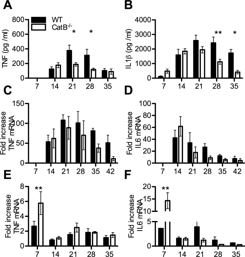
Fig 4. Innate immune cytokine responses of L. major infected WT and CatB-/- mice.
WT and CatB-/- were subcutaneously inoculated with 3x10⁶ stationary phase promastigotes of L. major in the footpads and experimental read-outs were measured weekly as indicated. Levels of TNF (A) and IL-1β (B) were measured in footpads of L. major -infected WT (filled bars) and CatB-/- (empty bars) mice by specific ELISA. Level of expression of TNF (C) and IL-6 (D) transcripts in footpads or TNF (E) and IL-6 (F) transcripts in lymph nodes of L. major -infected WT (filled bars) and CatB-/- (empty bars) mice were measured by RT-PCR. All PCR data values are normalized to the expression of the HPRT gene. Data depicted represent the mean and SEM of at least 3 independent experiments with n ≥ 4 mice/group/time point. * p<0.05; ** p<0.01; *** p<0.001; **** p<0.0001

