
Figure 4.
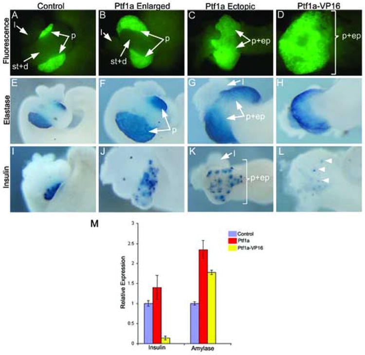
Overexpression of Ptf1a and Ptf1a-VP16 mRNA promotes ectopic and enlarged pancreas formation. Stage 42-44 dissected whole guts are shown in each image. (A) Control F2 Elas-GFP transgenic tadpole whole gut. (B) Ptf1a-injected embryos showing expanded GFP fluorescence. Stomach (st), duodenum (d) and liver (l) are normal, but the pancreas (p) is enlarged. (C) Ectopic pancreas formation in Ptf1a-injected embryo. The pancreas, stomach and duodenum (p+ep) form a large ectopic pancreas expressing Elas-GFP. (D) Ptf1a-VP16 injected embryo showing ectopic GFP fluorescence throughout the anterior endoderm. (E-H) In situ hybridization for elastase expression on isolated whole guts from (E) Control, (F,G) Ptf1a-injected embryos and (H) Ptf1a-VP16 injected embryos. Notice expanded and ectopic expression of elastase in Ptf1a and Ptf1a-VP16 injected embryos. (I-L) In situ hybridization for insulin in (I) Control, (J,K) Ptf1a-injected embryos and (L) Ptf1a-VP16 injected embryos. More insulin-expressing cells are detected in Ptf1a-injected whole guts, while there is a large decrease in insulin-expressing cells in Ptf1a-VP16 injected whole guts (arrowheads). (M) Real time PCR analysis of endocrine and exocrine markers in Ptf1a and Ptf1a-VP16 injected embryos for amylase and insulin expression in control, Ptf1a-injected and Ptf1a-VP16 injected whole embryos at stage 35. Each bar is an average of 4 individual whole tadpoles. Amylase expression is increased in both, while insulin expression is decreased in Ptf1a-VP16 and increased in Ptf1a-injected embryos. Purple-control tadpoles, red-Ptf1a mRNA injected tadpoles, yellow-Ptf1a-VP16 injected tadpoles.