
Figure 7.
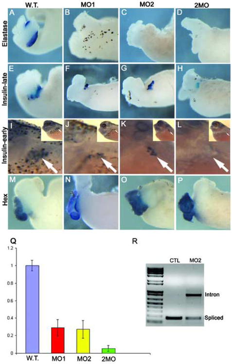
Elastase expression is absent in Ptf1a morphants, while insulin expression is reduced at late stages, but absent at early stages. (A-D) Whole mount in situ hybridization for elastase RNA expression in control and Ptf1a-MO dissected whole guts at stage 42. Elastase expression is not detected in either single or double morpholino injected guts. (E-H) Whole mount in situ hybridization for insulin RNA expression in control and Ptf1a-MO whole guts. (F,G) Insulin expression is decreased in both single morpholino injections. (H) In embryos injected with both morpholinos together insulin expression is also reduced, but still present. (I-L) Initial insulin expression is lacking in Ptf1a morphants. Insulin RNA expression by whole mount in situ hybridization in control and Ptf1a-MO injected embryos at stage 35. Inset in each panel is the low power view. (I) Control tadpole showing normal punctate insulin expression in the dorsal pancreas (arrow). (J,K) Insulin expression is reduced in single morpholino injected embryos, but (L) completely lacking in double morpholino injected embryos. (M-P) Expression of the liver marker Hex is normal in both single and double Ptf1a morpholino injected embryos. (Q) Real time PCR analysis of insulin expression in control and Ptf1a-MO injected embryos at stage 35 confirms insulin reduction in single and double morpholino injected embryos. Each bar is an average of four individual tadpoles. There is a 50-60% reduction in single morpholino tadpoles (40ng), but an almost complete absence in the double morpholino tadpoles (20ng each). (R) RT-PCR analysis of splicing in control and MO2-injected embryos showing the inhibition of splicing by MO2. Loading control was established with EF1α (not shown). Primers were designed flanking the single Ptf1a intron.