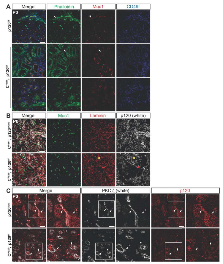
Figure 5. Changes in cytoskeletal architecture in pancreatic epithelium lacking p120 catenin.
Immunofluorescent staining of P0 pancreas showing the apical markers Muc1, Phalloidin, and PKCζ as well as the basal markers CD49f and Laminin in homozygous p120f/f and wild-type control pancreases. (A) Phalloidin was localized apically in wild-type ducts and apically, laterally, and basally in homozygous p120f/f ducts, indicating an alteration in cytoskeletal organization. Phalloidin was localized apically in the acini of both homozygous p120f/f and wild-type control pancreases. (A–B) Muc1 was localized apically in both wild-type and homozygous p120f/f pancreatic epithelium. White arrows point to Phalloidin and Muc1 staining in both wild-type and homozygous p120f/f ducts. Both basal markers CD49f and Laminin have comparable basal localization in wild-type and CPdx1; p120f/f pancreatic epithelium. (B) A section with unusually high mosaicism for p120 catenin was intentionally chosen to allow comparison of Muc1 and Laminin staining in p120 catenin-expressing and p120 catenin-deleted tissue in the same section. Yellow arrows point to a p120 catenin-expressing acinus and red arrows show a p120 catenin-deleted acinus. There was no difference in Laminin or Muc1 staining between p120 catenin-expressing and p120 catenin-deleted tissue. (C) In the wild-type panel, white arrows point to apical localization of PKCζ in ductal epithelium (left arrow) and in an acinus (right arrow). In the CPdx1; p120f/f panel, white arrows point to apical localization of PKCζ in an acinus (left arrow) and both apical and increased cytoplasmic localization of PKCζ in expanded ductal epithelium (right arrow). Higher magnification images are shown for each panel. Scale bars are 50μm.

