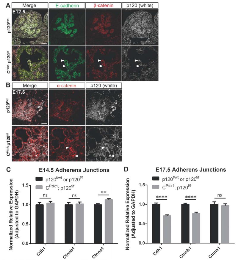
Figure 6. Adherens junction components are retained in p120 catenin null epithelia during development.
(A–B) Comparison of immunofluorescent staining of p120 catenin, E-cadherin, β-catenin, and α-catenin using embryonic tissues selected for the presence of mosaic p120 catenin-expressing epithelial cells revealed reduced levels but persistent presence of adherens junction components at cell membranes of epithelial cells lacking p120 catenin. White arrows point to a few p120 catenin-expressing epithelial cells in a largely p120 catenin-deleted pancreatic epithelium. Scale bars are 50μm. (C–D) Comparison of gene expression of Cdh1, Ctnnb1, and Ctnna1 between wild-type and homozygous p120f/f pancreata at E14.5 and E17.5 using qPCR. Note that expression of Cdh1 and Ctnnb1 was not significantly different at E14.5 but was reduced at E17.5 in homozygous p120f/f pancreases when compared to controls. Wild-type control targets were normalized to 1. For all genotypes and all genes at both E14.5 and E17.5 time points, n=7, and reactions were run in quadruplicate. For interpretation of statistical results from Student’s t test, ns = not significant and p value > 0.05, * = p value ≤ 0.05, ** = p value ≤ 0.01, *** = p value ≤ 0.001, and **** = p value ≤ 0.0001.

