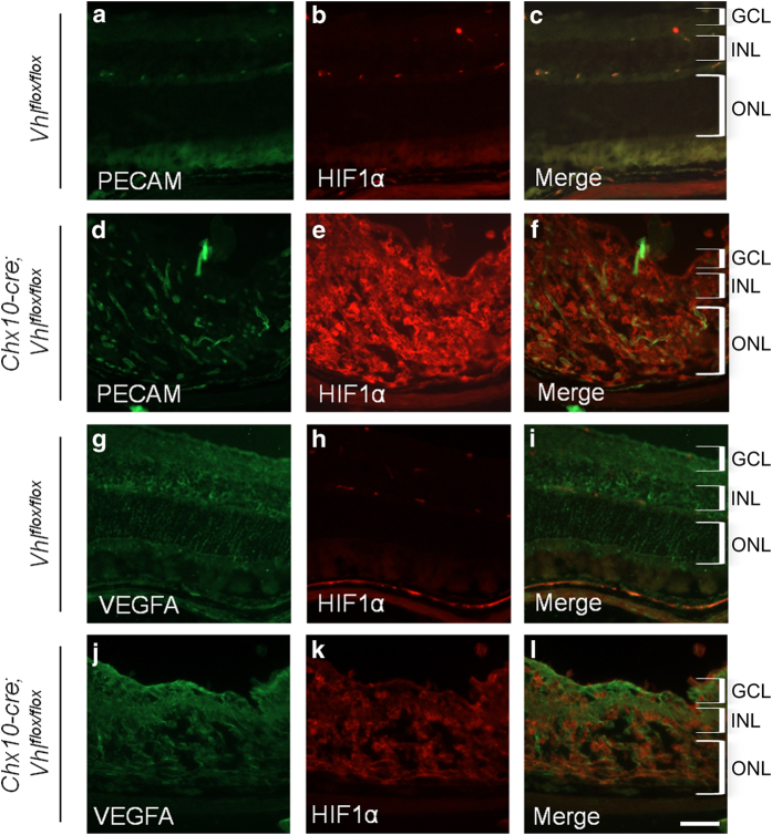
Figure 3.
Cre-mediated recombination causes increased HIF1α. Protein levels were analyzed by immunostaining frozen retinal sections for Vhlflox/flox cre-negative mice and Chx10-cre; Vhlflox/flox mutant mice at 1 month of age. Both PECAM (green; a) and HIF1α (red; b) protein levels were present in Vhlflox/flox cre-negative mice, but PECAM (d) and HIF1α (e) protein levels were only elevated after cre-mediated excision of the Vhl gene. Merged images for the Vhlflox/flox cre-negative mice (c) and Chx10-cre; Vhlflox/flox mutant mice (f) show combined PECAM (green) and HIF1α (red) protein levels in the retina. Vascular endothelial growth factor (VEGF; green) was similar between the Vhlflox/flox cre-negative mice (g) and Chx10-cre; Vhlflox/flox mutant mice (j), whereas HIF1α (red) was increased as expected in the mutant mice (k) compared with the cre-negative mice (h). Merged images for the cre-negative mice (i) and mutant mice (l) show VEGFA and HIF1α overlayed on the same figure. Both the sections for PECAM and HIF1α, and those for VEGFA and HIF1α were taken at the same exposure. GCL, ganglion cell layer; INL, inner nuclear layer; ONL, outer nuclear layer. Scale bar, 600 μm.

