Figure 7.
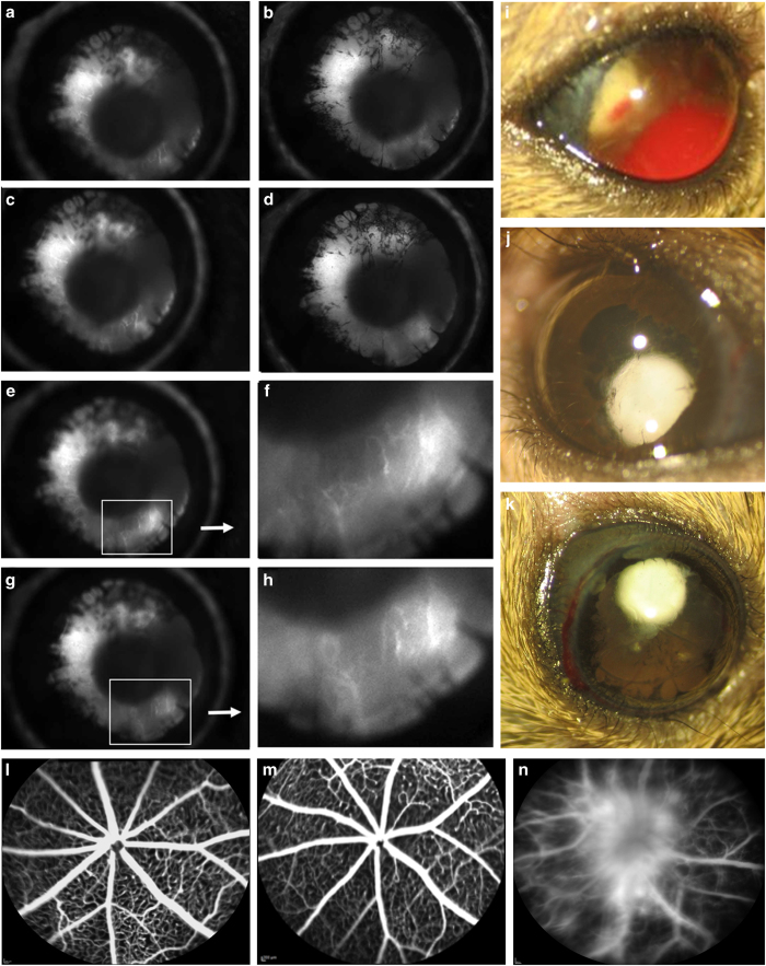
Neovascularization and leakage in the Chx10-cre; Vhlflox/flox mutant mouse mimics the human proliferative DR (PDR) phenotype. Time-lapse images from iris angiography at postnatal day 10 (P10) reveals anterior segment ischemia, rubeosis and iris synechaie (a–e, g). Enhanced images of fluorescein leakage in the iris during time-lapse angiography (f, h). t=10 s post-fluorescein dye injection. Fundus photographs of mutant mice at 3 months of age, showing visible vitreous hemorrhage (i), cataract (j) and signs of anterior segment ischemia (k). Fluorescein angiography in mice older than 1 month of age, for a wild-type C57BL/6J mouse (l) and a Chx10-cre; Vhlflox/+ heterozygote (m), uncovered no fluorescein leakage or detectable abnormalities of the retinal vasculature. In contrast, at P19, angiography of the Chx10-cre; Vhlflox/flox mutant mouse detected leaking fluorescein dye and capillary dropout, characteristic of PDR in humans (n). N⩾4 mice, phenotype was visible in all mice examined.

