Fig. 5.
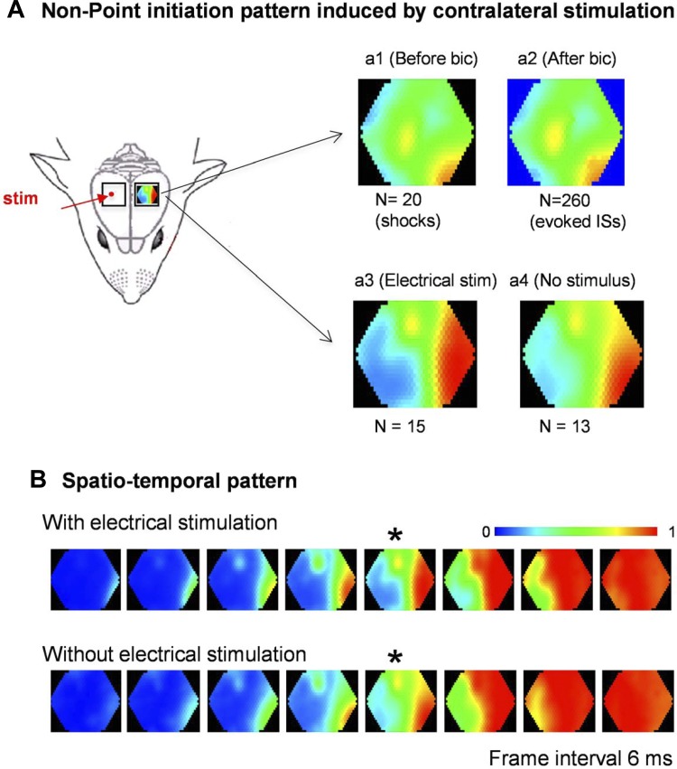
Point-stripe pattern induced by electrical stimulation to the contralateral cortex. A: contralateral point stimulation induced neuronal activities in the imaging side with a point-stripe pattern, both before (a1) and after (a2) bicuculline application. In another animal, the stimulation was turned off after bicuculline application to allow ISs to emerge without stimulation. Identical point-stripe initiation patterns are seen with (a3) and without electrical stimulation (a4). B: initiation patterns of the ISs with and without electrical stimulation. The images in a3 and a4 were selected from the frames marked by an asterisk (*).

