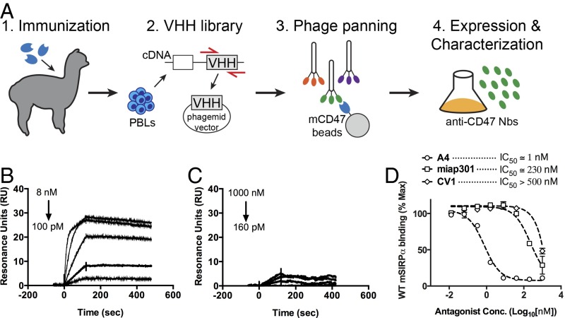
Fig. 1.
Characterization of anti-mouse CD47 antagonist nanobody. (A) Schematic depicting the generation of anti-mouse CD47 nanobody. An alpaca was immunized with the ECD of mouse CD47, and peripheral blood lymphocytes were isolated and used to create a camelid VHH phage library for selection of nanobodies that bind the mouse CD47 ECD. (B and C) Representative surface plasmon resonance (SPR) sensogram of anti-mouse CD47 nanobody A4 binding to immobilized mouse CD47 (B) or human CD47 (C). All sensograms were baseline-adjusted and reference cell-subtracted. (D) Dose–response curves of B16F10 cell surface CD47 antagonism with the anti-mouse CD47 nanobody A4, anti-mouse CD47 antibody miap301, and the anti-human CD47 antagonist CV1. Cells were incubated with increasing concentrations of CD47 antagonists and 100 nM fluorescent SIRPα tetramers for 1 h at 4 °C, washed, and evaluated by FACS. The data shown are the mean (n = 3), and error bars indicate SD. Dashed lines represent data fit to a one-site LogIC50 model in Prism.

