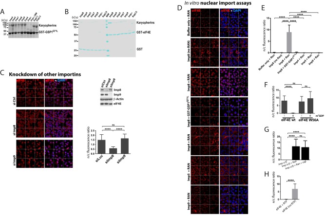
Fig. S1.
GST pull-down and in vitro nuclear import assays monitoring multiple karyopherins. (A) Activity of each karyopherin used in this study was verified by GST-GSP1 pull-down and visualized by Coomassie Blue (details are provided in SI Materials and Methods). (B) Direct interactions between different importins and eIF4E were examined by GST pull-down assay. The protein bands were visualized by Coomassie Blue staining after SDS/PAGE. The strongest target by far is importin 8 (Imp8). (C) Effects of knockdown of indicated importins on eIF4E localization in intact U2OS cells were investigated. Cells were stained with eIF4E antibody and DAPI as a nuclear marker. RNAi control or knockdown of given importins was tested as indicated. (Top right) Western blot analysis confirmed knockdown and that there were no effects on eIF4E levels. (Bottom right) Quantification is shown. (D) In vitro nuclear import assays using purified GST-eIF4E in digitonin-permeabilized U2OS cells in buffer (including transport cofactors, such as GTP, etc.) in the presence of importin 8 under various conditions and in the presence of other importins. For both C and D, confocal micrographs were single sections through the plane of the cells with a magnification of 63×. The confocal microscope collected nine fields (“tiles”) simultaneously and automatically reassembles these tiles to give a larger field. The grid depicts the borders for these tiles. Quantifications for confocal images are shown for Fig. S1D (E) and for Fig. 4 H (F) and G (G) and 5B (H). ****P < 0.0001. ns, not significant.

