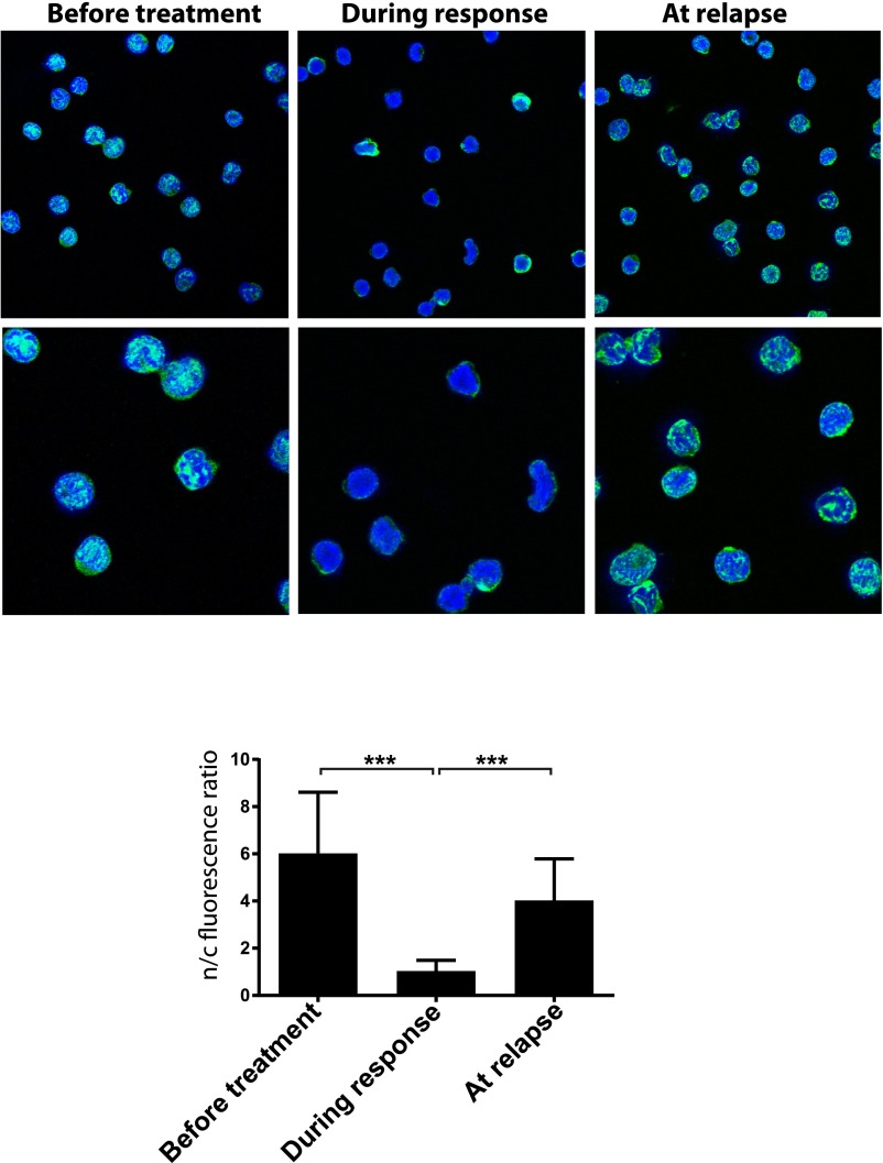
Fig. S10.
eIF4E localization is dysregulated in leukemia. eIF4E localization (green) in leukemia blasts cells isolated from a patient with AML treated with ribavirin [patient 6 in our ribavirin monotherapy trial (9)]. Confocal micrographs of patient 6 samples taken before treatment, during response, and at relapse. DAPI, a nuclear marker, is in blue. Micrographs are single sections through the plane of the cells. [Magnification: 100× (Top) with an additional 3× digital zoom (Bottom)]. Quantification of nuclear to cytoplasmic intensity ratio for eIF4E is shown in bar graph form. ***P < 0.001.

