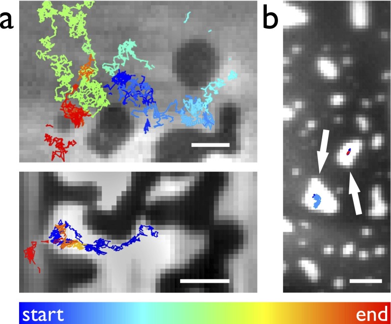
Fig. S6.
Electropore diffusion in phase-separated membranes. Median images of between 500 and 7,000 frames recorded at 99.4 Hz (A) or 49.9 Hz (B). Colored trajectories show motion of electropores; their color indicates the time at which the trajectory begins with respect to the start of the recording. (A) DPhPC/DPPG/cholesterol, 1:1:1. Electropores in the Ld phase diffuse around, but not within, areas of Lo lipid. The lower panel shows an electropore passing between a gap in the ordered region. (B) In a DIB formed of DPhPC/bSM/cholesterol, 1:1:1, electropores form and are trapped within the small regions of the Ld phase, marked by arrows. (Scale bars: 5 μm.)

