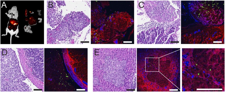
Fig. 4.
Targeted detection of orthotopic ovarian tumors. (A) NIR-II images of the whole mouse (left) and the excised organs (from left to right and top to bottom are, spleen, ovaries, kidneys, pancreas, liver, stomach, and intestines). The bright spots indicate tumor nodules. (B–E) Slices of tumor (B), tumor nodules on pancreas (C), intestine (D), and the tumor invaded the liver (E). Each pair show an H&E-stained slice of tissue (Left) and the registered multiphoton confocal microscopy (Right) where signals from DCNPs (green), hematoxylin (blue), and eosin (red) were composited. (Scale bars: 100 μm.)

