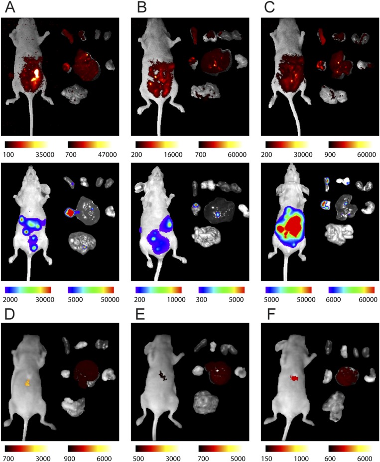
Fig. S5.
Targeted detection of orthotopic ovarian tumors by i.p. administration of LbL DCNPs. (A–C, Top) NIR-II fluorescence images of tumor-bearing mice (Left) and excised organs (Right) at 72 h postinjection. (Bottom) bioluminescence images of the same tumor-bearing mice (Left) and excised organs (right) at 72 h postinjection. (D–F) NIR-II fluorescence images of tumor-free mice (Left) and excised organs (Right) at 72 h postinjection. For the excised organs, from left to right and top to bottom, organs are presented in the order of spleen, ovaries, kidneys, pancreas, liver, stomach, and intestines. Color bars show the low- and high-intensity values in units of counts/s.

