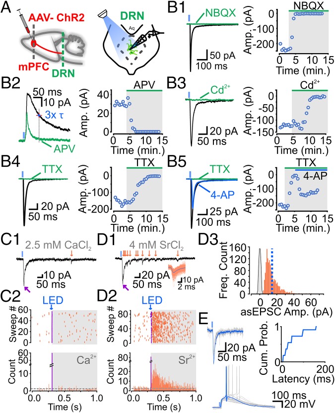
Fig. 1.
mPFC-DRN inputs onto 5-HT neurons are monosynaptic, glutamatergic, and sufficient to drive action potential firing. (A) Schematics of adeno-associated virus (AAV)-ChR2 injection (Left) and DRN neuron recording from a midbrain slice (Right). ChR2-PSCs are blocked by NBQX (10 μM) or D-APV (50 μM) (B, 1: n = 5; B, 2: n = 5). Presynaptic release is inhibited by cadmium (Cd2+; 200 μM) or TTX (1 μM) (B, 3: n = 3; B, 4: n = 10). (B, 5) Inhibition of presynaptic release with TTX was partially rescued by 4-AP (1 mM; n = 6). Traces of a ChR2-EPSC (magenta arrow) and spontaneous EPSCs (sEPSCs) (orange arrows) in the presence of 2.5 mM extracellular Ca2+ (C, 1) or 4 mM strontium chloride (SrCl2) (D, 1) are shown. (C, 2, Top and D, 2, Top) Scatter plot of ChR2-EPSCs (magenta) followed by sEPSCs (orange). (C, 2, Bottom and D, 2, Bottom) Peristimulus time histogram of ChR2-EPSCs (magenta) and sEPSCs (orange). (D, 3) Histogram of asynchronous EPSC (asEPSC) amplitudes at mPFC-DRN synapses onto 5-HT neurons (n = 9). (E) Recordings of ChR2-EPSCs [Left, membrane voltage (Vm) = −70 mV] and action-potential firing in current-clamp recordings (Right, Vm ∼ −70 mV). Cumulative distribution plot depicts the latency distribution of mPFC, i.e., mPFC-driven action potentials in DRN 5-HT neurons (49.4 ± 14.5 ms, n = 11). Amp., amplitude; Cum. Prob., cumulative probability; Freq., frequency.

