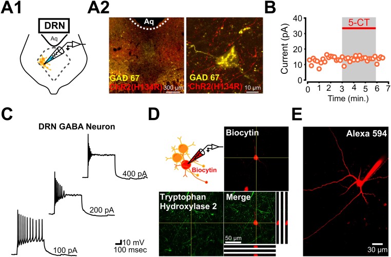
Fig. S6.
Electrophysiological and immunohistochemical identification of local GABAergic interneurons located in the lateral wings of the DRN. (A1) Schematic of whole-cell recording from a DRN GABAergic interneuron. (A2) Immunostaining for GAD67 and ChR2(H134R)-mCherry. (Right) Higher magnification image of a DRN GABAergic neuron surrounded by ChR2-expressing mPFC axons and terminals. (B) Whole-cell voltage-clamp recording from a DRN GABA neuron with no significant change in holding current in response to bath application 5-CT (Vm = −55 mV). (C) Characteristic firing properties of a DRN GABAergic interneuron in the DRN. (D) Post hoc immunolabeling for TPH2 in a GABAergic interneuron filled with biocytin during a whole-cell recording. TPH2 was not detectable in the GABAergic interneuron. (E) Two-photon image of Alexa-594–filled GABAergic interneuron in the DRN. Note the extent of the dendritic arborization of the DRN GABAergic interneuron.

