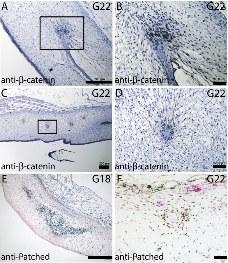
Fig. S2.
Osteogenic signaling in developing plastron bones and spicules. (A–D) Nuclear β-catenin staining of the cells at the proliferative osteogenic front of plastron bone (A and B) and surrounding bone spicules at G22 (C and D). (E) G18 transverse sections stained with anti-Patched antibody indicate Hedgehog signaling in the preosteoblastic cells in plastron bone, and (F) surrounding bone spicules at G22. [Scale bars, ∼200 μm (A, C, and E) and 50 μm (B, D, and F).]

