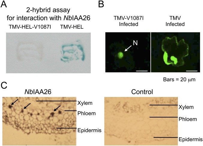
Fig. S3.
Characterization of NbIAA26. (A) Yeast two-hybrid assay showing the interaction of the TMV-helicase-bait protein and NbIAA26-prey protein and the absence of interaction between TMV-V1087I-helicase-bait protein and NbIAA26-prey protein. (B) TMV infection interferes in the nuclear localization of NbIAA26-GFP. Fluorescent images of N. benthamiana leaf tissue transiently expressing NbIAA26-GFP fusion protein in TMV-V1087I–infected or TMV-infected cells. Yeast two-hybrid and transient localization assays were done as previously described (20). (Scale bars, 20 μm.) (C) In situ immunolocalization of NbIAA26 in N. benthamiana stem cross-sections. Dark brown color indicates NbIAA26 mRNA accumulation. Arrows denote phloem location.

